After-hours emergencies (612) 555-0199

Meridian OralOral and maxillofacial surgeryReferral form
Reading as

Procedures, imaging requirements and the referral form.

Plain language, preparation and recovery instructions.

Surgery a referring dentist can hand over without worrying.

You have been referred. Here is exactly what happens next.

Classification, canal relationship and a written plan before your patient is seen. We place; you restore.

Being sent to a specialist is a normal step, not a sign that something has gone wrong. This explains what happens, how it will feel, and when to call.

Referral triage
Same day
Surgeons
Three
On-site imaging
CBCT and IOS
Plate 01 — right hemimandible, lateral aspectA line drawing of the right half of a lower jaw seen from the side. The premolar and two molars stand in the bone. A wisdom tooth behind them is tipped forward at about forty-five degrees. An amber line traces the inferior alveolar canal from the mandibular foramen in the ramus, forward beneath the tooth roots, to the mental foramen below the premolars. Fine dashed guide lines and measurement ticks are drawn around the bone.CondyleCoronoidM3 · mesioangularIAN canalMental foramenAlveolar crest
Plate 01 · right hemimandible, lateral aspectScale 1 mm = 3.5 units · drawn, not traced
MeasurePick a point

The switch in the header re-tabs the whole site.

A referring dentist and a frightened patient want opposite things from the same sentence, so the control at the top changes the navigation, the register that leads, and the order of the columns in the pathway below.

Reading as a dentistClassification, canal relationship, technique, turnaround, what comes back to you.
Reading as a patientWhat will happen, how long it takes, what it will feel like, and when to call.
NeverHidden. Clinical detail is not withheld from patients, and plain language is not withheld from clinicians. The switch decides which you meet first.

Plates 01 to 07

Seven areas, drawn in section.

Hover, focus or tap a block and it is sectioned: the cover plate hinges open on its cut plane, the thickness of the cut shows on the exposed edge, and the interior layers stand at their own depths.

Fig 01 Mucosa & flapSagittal
Plate 01Third molar removalAssessment and removal of impacted and partially erupted third molars, including coronectomy where the root is intimately related to the inferior alveolar canal.Taking out wisdom teeth that are stuck, coming through crooked, or causing repeated infections — and knowing when it is safer to leave part of a root alone.20–60 minRead the plate
Fig 02 Gingiva & papillaeCoronal
Plate 02Dental implantsSingle-tooth and partially edentulous implant placement, immediate or delayed, planned from a CBCT against a stated restorative endpoint.A titanium root placed into the jawbone, which your own dentist later builds a tooth onto. It is two appointments months apart, not one.45–90 min per siteRead the plate
Fig 03 Mucosa & prosthesisSagittal
Plate 03Full-arch restorationFixed full-arch rehabilitation on four to six implants, with tilted posterior fixtures where anatomy allows, and immediate provisionalisation where cross-arch stability permits.A fixed set of teeth for a whole jaw, held on four to six implants. It is a big decision, it is not reversible, and it needs cleaning every day for the rest of your life.3–5 hoursRead the plate
Fig 04 Flap & membraneCoronal
Plate 04Bone & soft-tissue graftingRidge preservation, guided bone regeneration, lateral-window and transcrestal sinus augmentation, and keratinised-tissue grafting around teeth and implants.Rebuilding bone or gum where there is not enough of either — usually so an implant can be placed later, and usually months before it.45–120 minRead the plate
Fig 05 Soft-tissue profileSagittal
Plate 05Corrective jaw surgeryOrthognathic surgery — Le Fort I, bilateral sagittal split osteotomy, genioplasty and SARPE — planned virtually and phased with orthodontic decompensation.Surgery to move the upper jaw, the lower jaw or both, so the teeth meet properly and the face is balanced. It runs alongside two to three years of braces.2–5 hoursRead the plate
Fig 06 Capsule & lateral pterygoidCoronal
Plate 06TMJ & facial painAssessment of temporomandibular disorders, with conservative management first and staged intervention — arthrocentesis, arthroscopy, open arthroplasty — reserved for defined failures.Jaw joint pain, clicking and locking. Most of it settles with treatment that does not involve surgery, and that is where we start.Consultation 45 minRead the plate
Fig 07 Mucosa & lesionSagittal
Plate 07Oral pathology & biopsyAssessment and biopsy of mucosal and intra-osseous lesions, with a same-week pathway for anything suspicious and direct referral into head-and-neck oncology where required.Getting a definite answer about a lump, patch or ulcer in the mouth by taking a small sample and having a pathologist look at it.20–40 minRead the plate

Referral to recovery, in five steps.

The clinical column and the plain-language column say the same thing to two different readers. Your choice at the top decides which one leads.

Referral · Same day

The referral arrives

Clinical

  • Secure portal, encrypted email or fax — whichever your practice already uses.
  • Triaged the day it arrives; anything with a red flag is pulled out of the queue.
  • Imaging reviewed before the appointment is offered, so the slot length matches the case.
  • A written acknowledgement back to you with the triage decision.

In plain language

  • Your dentist sends us your details, your x-rays and what they are worried about.
  • We look at the x-rays before we offer you a time, so the appointment is the right length.
  • You will hear from us to book. If it is urgent we will call rather than write.
Consultation · Within 1–3 weeks

Assessment and consent

Clinical

  • Full medical history including anticoagulants, antiplatelets, antiresorptives and antiangiogenics.
  • ASA classification recorded and anaesthesia planned against it.
  • Examination, imaging review, and a written plan with named risks.
  • A copy of the consultation letter to you the same week.

In plain language

  • We examine you, look at the scans with you, and explain what we think should happen.
  • Bring a list of every medicine you take, including ones from the hospital and anything you buy yourself.
  • You will hear the risks out loud, not just read them. Ask us anything, twice if you like.
  • Nothing is booked on the day unless you want it to be.
Imaging · Same visit where possible

Imaging and planning

Clinical

  • CBCT on site with a field of view matched to the question being asked.
  • Intraoral scanning for implant and orthognathic planning.
  • Virtual planning for full-arch and orthognathic cases, shared before the surgical date.
  • DICOM available to you on request — we would rather you had the data than a screenshot.

In plain language

  • Most scans happen here, usually on the same visit, and take a couple of minutes standing still.
  • A cone beam scan is a 3D x-ray. The dose is higher than a normal dental x-ray and much lower than a hospital CT.
  • We only take one if it will change what we do.
Surgery · Scheduled to the case

The procedure

Clinical

  • Office-based under local, local with IV sedation, or general anaesthesia; hospital for orthognathic and major reconstruction.
  • Antibiotic prophylaxis by indication, not by habit.
  • Anticoagulation managed to current guidance — we do not routinely stop DOACs for simple dentoalveolar surgery.
  • Operative note and post-operative regime to you within 24 hours.

In plain language

  • You choose, with us, how awake you want to be. Sedation means you will not remember much of it.
  • If you are having sedation you need an adult to bring you and take you home, and to stay with you.
  • You will leave with written instructions and a number to call. Please actually take the written instructions.
Recovery · Days to months

Recovery and handback

Clinical

  • Review at 7–14 days for most cases; earlier where there is a specific concern.
  • Discharge letter with the histology, the fixture record or the fixation detail, as applicable.
  • The patient returns to you for restorative care — we place, you restore.
  • Direct line stays open to you for as long as the case needs it.

In plain language

  • Swelling peaks around two to three days and then improves. That is normal and expected.
  • The recovery page tells you what to eat, what to expect, and exactly when to call.
  • You go back to your own dentist for the rest of your care.

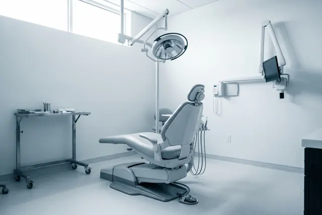
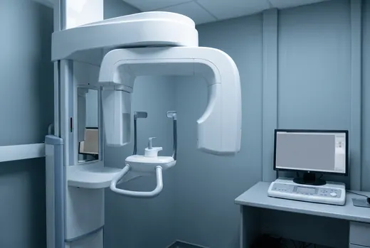
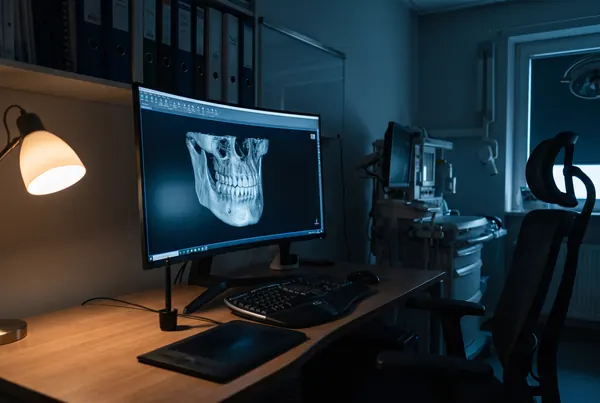
Three operatories, one imaging suite.

An empty, prepared oral surgery operatory with an articulated surgical light and a stainless bracket table
Office surgical suite, preparedA
A cone beam CT scanner in a lead-lined dental imaging room with its control console
CBCT, small field of view by defaultB
A planning workstation showing a three-dimensional reconstruction of a jaw in blue wireframe
Virtual planning, shared before the dateC

Anaesthesia

Local, local with intravenous sedation, or general anaesthesia. The permit is held by Dr. Adeyemi, monitoring is continuous, and the emergency drugs and airway equipment are checked to a written schedule before every sedation list.

How anaesthesia is decided

What we send back

An operative note and post-operative regime within 24 hours. For implants, the fixture record with platform, torque and a date the site is ready to impress. For biopsies, the histology report the day it lands.

The referring doctors page

Two kinds of evidence, kept apart.

Notes from referring practices describe how a case was handled. Patient accounts describe how a recovery felt, anchored to a day count. Different claims, so they are not averaged into a score, and there is no star rating anywhere on this site.

Third molar
The report came back before the patient did. Classification, canal relationship, and a clear recommendation for coronectomy with the reasoning attached — I could answer her questions at her hygiene visit without guessing.
Dr. R. OkonjoLyndale Family Dental, Minneapolis, MNReferred: Impacted 38, canal contact on CBCT
Implant
They send a fixture record with the platform, the torque and a date the site is ready. I have stopped having to telephone to find out what was placed.
Dr. S. PelletierNokomis Dental Group, Minneapolis, MNReferred: Implant 46, delayed placement
Pathology
I sent a lesion on a Thursday that I was not comfortable watching. It was seen the following Tuesday and I had the histology report the week after. That pathway is the reason they get everything urgent I have.
Dr. M. HollanderUptown Dental Care, Minneapolis, MNReferred: Non-homogeneous leukoplakia, lateral tongue
TMJ
They declined to operate on one of my TMD patients and sent back an exercise programme and a note explaining why. I was mildly annoyed and entirely wrong — she was better in six weeks.
Dr. A. FreeburgSt. Anthony Dental, St. Paul, MNReferred: Arthralgia, three months duration
Orthognathic
Our orthognathic case was planned jointly from the first appointment. The planned movements were shared with me before the theatre date and the post-surgical detailing was exactly what the plan predicted.
Dr. J. WhitfieldWhitfield Orthodontics, Edina, MNReferred: Skeletal III, BSSO setback with Le Fort I
Grafting
The bit I value most is being told no. When a graft was not going to be predictable they said so and offered a shorter fixture instead, rather than selling me a bigger operation.
Dr. P. NakamuraCedar Riverside Dental, Minneapolis, MNReferred: Vertical deficiency, posterior mandible
Record 1 of 6

Notes are reproduced with permission from the referring practice and edited only for length. They describe how a case was handled, not how it turned out. This practice publishes no outcome claims.

The after-hours line is answered by a surgeon.

Not a call centre, not a message service. One of the three of us, on the phone, tonight.

Send us a case.

Ask us anything before your appointment.

The referral form takes about two minutes. If it is urgent, do not use the form. Call the referral line on (612) 555-0150 and you will reach a surgeon.

You do not need to know the right words. Tell us what you have been told and what you are worried about, and we will take it from there.

Open the referral form Request an appointment

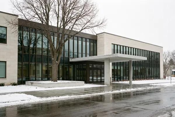
The low-rise limestone and bronze medical building housing the practice, photographed on an overcast winter day
2100 Hennepin Avenue, Suite 400, MinneapolisD